Obfuscated pancakes

Anything is better than an recipe that uses volume measurements for highly compressible powders. I’m looking at you America, and your ridiculous ‘cup’ nonsense.

Materials
  • 0.110 kg powdered wheat endosperm (without added carbonates or tartrates)
  • 4.28 mmol NaCl
  • 2 unfertilised jungle-fowl eggs (between 63 and 73 g in mass)
  • 275 mL of 75% (v/v) aqueous diluent of cow’s milk
  • 45.0 cm3 cow’s milk butter
  • 1 hesperidium from a lemon-tree, cut into ungulae with semidisc dihedrals of π/2
  • 55.8 mmol granular α-D-glucopyranosyl-(1→2)-β-D-fructofuranoside (particle size c. 350 microns)
Method

Inorganic materials were dissolved into the diluted milk. Shells and chalazae of the eggs were discarded and the remaining components were thoroughly combined with the endosperm, diluted milk, and 40% of the mass of the butter (warmed until fused). A frying-pan was heated to 500 K using a methane flame and lubricated with one eighth of the remaining butter. Sufficient batter mixture was added to the pan to form a disc of radius c. 70 mm and thickness c. 0.25 mm. The under-surface was maillarded to the point of melanoidinisation (c. 0.5 min). The disc was then rotated half a turn about a randomly chosen diameter, and heated for a further 0.1 min. Discs were served with the granulated furanoside and berry portions.

Illuminating error

Yesterday’s first-year biology practical involved forcing the laboratory work-horse bacterium Escherichia coli to take up a circular piece of DNA called pGLO.

pGLO contains a few genes, but the most interesting of these DNA sequences encodes a protein from a jellyfish. This protein fluoresces green under UV light, and goes by the thoroughly unimaginative name of green fluorescent protein (GFP). The production of the GFP is regulated by a second DNA sequence, this time taken from a bacterium. The normal job of this regulatory DNA sequence is to switch on and off the production of enzymes that degrade an uncommon sugar called arabinose. Since bacteria don’t often encounter arabinose, the enzymes are not produced unless there is actually some arabinose around to eat. This save resources.

The system on the pGLO is therefore quite artificial: it patches together regulatory DNA from the arabinose-degrading genes, with protein-coding DNA from the jellyfish. The advantage of this is that arabinose will switch on the production of the GFP, which is much easier to see than the arabinose-degrading enzymes.

So the green, fluorescing bits in the plates below show where the bacteria are detecting arabinose and being tricked into making GFP.

pGLO contamination [CC-BY-SA-3.0 Steve Cook]The only thing is, they shouldn’t be! The plates these bacteria are growing on do indeed contain arabinose, as you might expect, but they also contain lots of glucose. Arabinose is very much second-best as far as the bacteria are concerned: they would much prefer to eat glucose than arabinose. If there’s glucose present as well as arabinose, the regulatory DNA sequence should switch off the production of the arabinose-degrading enzymes. In these bacteria with pGLO, the glucose should therefore switch off the production of GFP, and this plate should be dark as pitch.

The reason for the unexpected glowing bits is interesting. If you squint, you’ll see that there are four or five fluffy things on the plate as well as the glowing bacteria. These were not supposed to be there. They are fungal colonies, probably of Penicillium; contaminants that have gotten into the plates from the air, possibly ferried into the lab on the clothes of an unsuspecting undergraduate with a fridgeful of mouldy bread.

As the fungi have grown across the plates, half-smothering the bacteria, they have gobbled up all the glucose. The bacteria, starved of their preferred sugar, respond by switching on their arabinose-degrading enzymes and any other genes that happen to be controlled by that same regulatory DNA. And they glow.

Happy accidents are often more illuminating than the “expected results” from practicals that “work”.

Organism of the week #5 – Something’s wrong here

Busy teaching this week, so it’ll have to be a quick one…

Drosophyllum lusitanicum [CC-BY-2.0 Alex Lomas]

Drosophyllum lusitanicum, Portuguese “sundew”

I particularly like this plant because of a very specific and unusual thing about its leaves. The more observant of you may spot what it is without having to resort to Wikipedia…

Organism of the week #4 – Toadstools

Imperial’s campus in Berkshire, Silwood Park, is a fabulous place to go fungus spotting. The fly agaric (Amanita muscaria) is very common there as there are a lot of birch trees around, and this fungus forms a symbiosis with the roots of those trees:

Amanita muscaria [CC-BY-SA-3.0 Steve Cook]Fly agarics are rather poisonous (for some value of ‘rather’), by virtue of containing muscimol, and ibotenic acid; the former is a psychoactive dissociative that can cause visual and auditory hallucinations. There is an enormous amount of folklore (i.e. lies) associated with this mushroom that I shan’t bother repeating. There’s not even much evidence that it’s any use for killing flies.

Although fly agarics are toxic, they are nothing like as poisonous as their amanitin-containing relatives, the cheerily named death cap (Amanita phalloides) and the delightfully named destroying angel (Amanita virosa), whose toxins don’t merely bugger up your nerves, but poison every cell in your body at an intimate molecular level. Fungi probably contain toxic compounds for the same reason as many plants and corals do: even if you can’t bite or run away from your predators, you can still make them very, very sorry.

Organism of the week #3 – Switch off all electrical items on the approach

There are many good reasons to visit San Francisco, some of them thoroughly unsafe for work, but one I didn’t consider was to see this amazing sight on the approach to the airport:

Halobacterium salinarum [CC-By-SA-3.0 Steve Cook]

Salt ponds at the southern end of San Francisco Bay

The pink colour in the water is caused by a bloom of single-celled organisms I must refrain from calling bacteria, since they are not. Nor are they eukaryotes, the group to which you and I, and plants and fungi and protists belong. The things living in the water are halo’bacteria’, named before it was realised we needed a whole new classificatory scheme to accommodate their weirdness. Halobacterium is a member of the Archaea, which includes many extremophiles. Some are capable of living in near-boiling water, hot acid, or – as here – in very salty water indeed, but some are much more commonplace and allow some humans to set light to their farts.

As well as being not-bacteria and not-eukaryotes, halobacteria also confound naïve human assumptions about how to make a living. If you look only at multicellular organisms, you find they fall mostly into two camps: photosynthetic organisms like plants, which need light, water, carbon dioxide, and not much else to grow; and heterotrophic organisms like humans and fungi, which steal almost everything they need to grow either directly or indirectly from photosynthetic organisms.

Halobacteria are pink because they contain a pigment called bacteriorhodopsin. They use this pigment to capture light from the Sun. This energy is used to run their biochemistry and to help pump salt into the cell to prevent it from popping from osmotic stress. But unlike green plants, they can’t fix carbon dioxide into sugars and other complex organic compounds. Like us, they have to get their carbon in a fixed organic form, mostly in the form of amino acids. Many bacteria and archaea have metabolisms that don’t fit the dreary ‘plant-like’ vs. ‘animal-like’ division, and this is one of the many reasons I love them so.

More butterworts

After the trip to Königssee, I was inspired to retail therapy and bought myself as many butterworts as Hampshire Carnivorous Plants could supply:

Butterwort collection [CC-BY-SA-3.0 Steve Cook]

OCPD butterwort collection (Pinguicula cv. Tina, P. cv. Wesser, P. agnata, P. cyclosecta, P. esseriana)

They did me proud over this summer, with two of them flowering despite the root disturbance (on which butterworts are not keen), the chunky cultivar ‘Tina’:

Pinguicula cv. Tina flowers [CC-BY-SA-3.0 Steve Cook]

Pinguicula cv. Tina flowering

and the daintier species P. esseriana:

Pinguicula esseriana flowers [CC-BY-SA-3.0 Steve Cook]

Pinguicula esseriana

In winter, a lot of butterwort species give up on making large, sticky, carnivorous leaves, and instead produce compact non-carnivorous rosettes that look a bit like houseleeks:

Pinguicula cyclosecta winter rosette [CC-BY-SA-3.0 Steve Cook]

Pinguicula cyclosecta winter rosette, remains of carnivorous summer rosette slowly composting away

Carnivory is not free, so this is probably an adaptation to the lack of insect prey available during the winter months.

When I grow up I want one of these.

I gone done made me a satire

Tortured artists have tuberculosis, not iPhone apps.

Instant Hipstagrammatic [CC-BY-SA-3.0 Mark Zuckerberg, I mean Steve Cook]

Farewell Marlowe

Marlowe, the cellar spider who has been living behind my beside cabinet for the last 18 months, disappeared a few weeks ago, but I wasn’t too worried as they’d done that before and then turned up again a few weeks later, fatter and of greater span.

On Monday, however, Alex found Marlowe’s mortal remains beneath the bathroom radiator. Marlowe is no more.

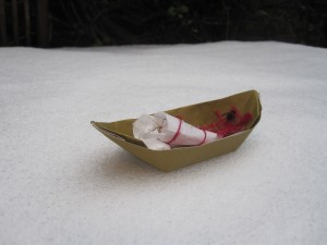
Rather than an undignified flushing or binning, I thought an undignified burning was in order, given the Viking weather the UK is currently experiencing:

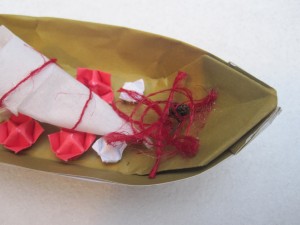
Marlowe's longboat (CC-BY-SA-3.0 Steve Cook)Marlowe was well-supplied with grave goods – origami flowers, a votive offering of a dead fly, and a ceremonial hemp web – for their journey we know not whither:

Marlowe's grave goods (CC-BY-SA-3.0 Steve Cook)

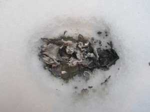
Purifying fire released their elements back to the air from which they came (somewhat indirectly, being a secondary consumer):

Marlowe's funeral pyre (CC-BY-SA-3.0 Steve Cook)Ashes to ashes, dust to dust.

Marlowe ashes to ashes (CC-BY-SA-3.0 Steve Cook)

Farewell Marlowe. May your spiderlings inherit the Earth.


Organism of the week #2 – Now wash your hands

I have delusions of being a competent recogniser of things that turn up in UK gardens, but when this started growing in what had previously been a pot of basil in the garden, I was clueless:

Phycomyces blakesleeanus [CC-BY-SA-3.0 Steve Cook]

Mystery thing

It looked a little like moss from a distance, but the bibbly bobblies were most certainly not moss-like close-up, and the whole thing had a rather unpleasant squeaky texture. Touching it left brownish-green stains on my fingers. Definitely not a moss.

A clue to its identity came when I unwisely tried poking about looking for roots, and realised it was growing on a cat turd.

Having suppressed my nausea and thoroughly washed my hands, a quiet bell from the mycology lectures I took during the Bronze Age rang. A vague memory of pin-moulds, big as your head, grasping for the light.

And indeed that is what this was. Phycomyces blakesleeanus is a giant relative of the tiny pin-moulds that grow on damp bread. A welcome addition to an otherwise lifeless and drab winter garden.

Organism of the week #1 – Chimaera

I’ve been neglecting the blog of late, for reasons too dreary to explain. So I am taking the astonishingly original step of racking up a load of pretty pictures of organisms to release on a semi-regular basis. I am creativity incarnate.

Today, +Laburnocytisus ‘Adamii’.

+Laburnocytisus adamii [CC-BY-2.0 Alex Lomas]

+Laburnocytisus ‘Adamii

Technically, this is a bit of a cheat for an organism of the week, as this organism is technically two organisms, one growing inside of the other.

This pretty mess is the result of a horticultural grafting going wrong. In a normal grafting, a bud from one plant (the scion) is inserted into a cleft cut into the stem of another (the rootstock). If the grafting behaves itself, you will then end up with the branches and fruit of one tree growing on the roots and trunk of another. Grafting is crucial in the farming of apples. If you plant the seeds from a nice apple, like an Egremont Russet, the resulting trees will take many years to get large enough to produce what will almost certainly be terribly disappointing fruit: most cultivated species of apple do not come true from their seed. Instead they have to be propagated vegetatively, and this is where grafting comes in. By grafting scions from an Egremont Russet onto a mass-produced rootstock (whose fruit you care not one jot about, but which has fabulous dwarfing roots), you can have an productive orchard making pleasant fruits within a few years, rather than the decades and more that would be needed to create a(n un)lucky-dip orchard from seed.

Grafting works best between closely related species. When it doesn’t work properly, the usual result is a dead scion. However, sometimes grafting goes just a bit wrong, and that is what has happened with +Laburnocytisus. Rather than having a scion growing on a rootstock separated by a scar, the scion and rootstock tissues have become mixed up in the scar, and are attempting to grow together. These somewhat unhappy marriages are called graft chimaeras.

The core tissue of +Laburnocytisus is Laburnum anagyroides, a popular child-killing shrub, which goes by the common name of ‘golden showers’ (*titters*). Growing over this core is a skin of tissue from another member of the bean family, the purple broom (Chamaecytisus purpureus).

Laburnum and broom are closely related, so for the most part, the two tissues manage to communicate their intentions quite well, and the leaves and flowers they produce are intermediate between the two species. But sometimes the laburnum core grows more quickly than the broom skin, and a chunk of laburnum bursts out in a shower of golden flowers, as you can see in the photo above.

Load more